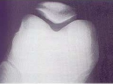
TANGENTIAL PATELLA PROJECTION
Axial • Modified Merchant View • Evaluation of patellar articular surface
SPECIALIZED PROJECTION
Specific projection for the evaluation of the patellar articular surface
Exposure Factors
Equipment: With bucky. Position: Prone.
Cassette Size
Visible Anatomical Structures
Complete Patella
Articular surface and body
Patellofemoral Space
Patellofemoral joint
Femoral Condyles
Lateral and medial
Joint
Proximal tibiofibular
- Articular surface of the femur - Femoral trochlea
- Lateral condyle of the femur and tibia - External compartment
- Medial condyle of the femur and tibia - Internal compartment
- Articular surface of the patella - Posterior patellar face
- Articular surface of the femur - Anterior femoral face
- Patella-femur relationship - Patellofemoral alignment
- Intercondylar space - Intercondylar fossa
- Articular cartilage - Patellofemoral space
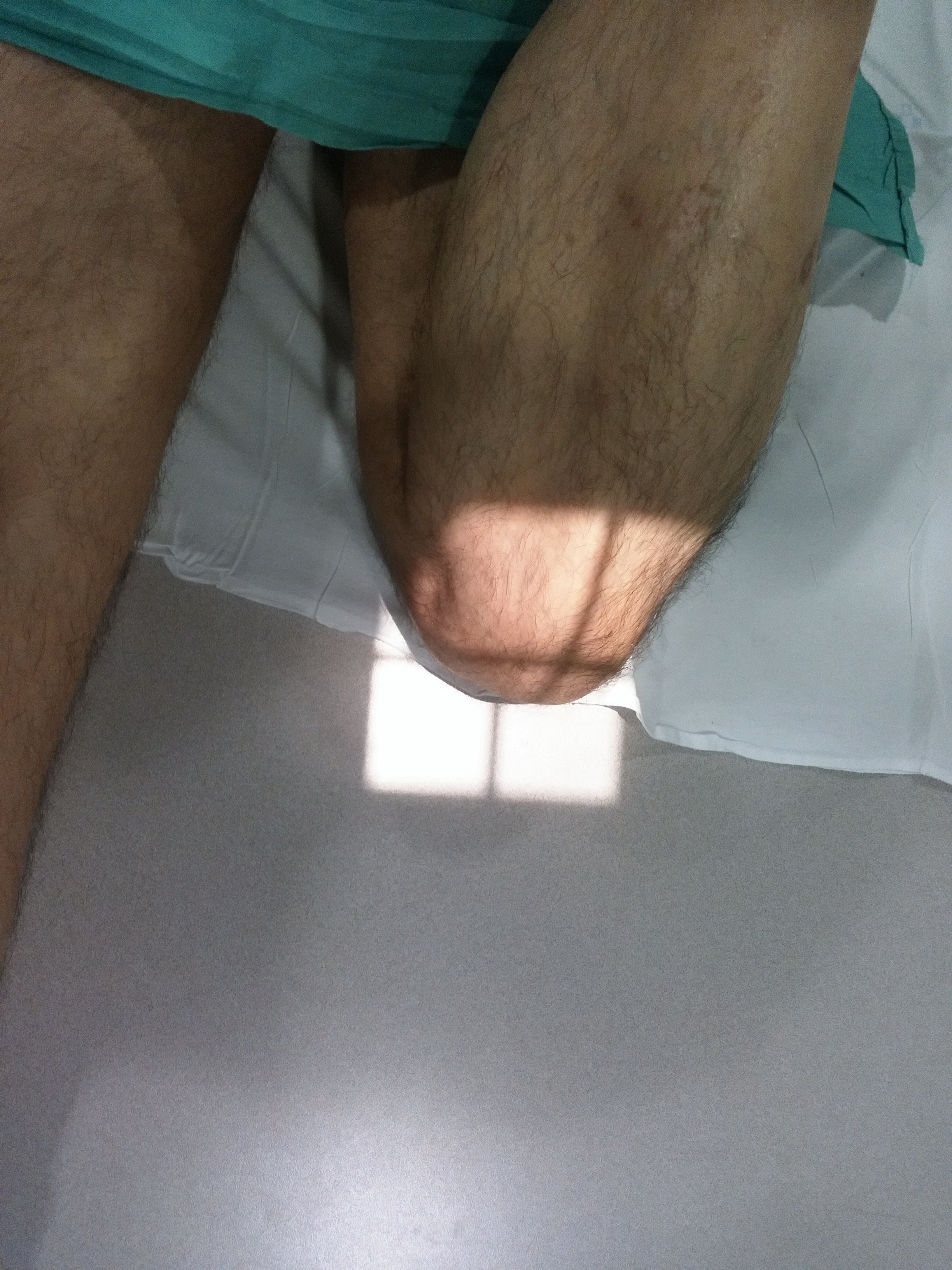
MANUAL FLEXION TECHNIQUE
Knee flexion is achieved through manual traction using a bandage or cloth that the patient pulls in a cranial direction during the examination.
This allows the patella to remain perpendicular to the table plane while the knee is flexed.
Patient Positioning
CENTERING POINT
Center on the patellofemoral joint for optimal visualization
Central Ray Direction
Vertical directed to the patellofemoral space
Trajectory: Perpendicular to the cassette
Entry point: Posterior, at the level of the intercondylar space
Exit point: Anterior, patellofemoral space
Purpose: Tangential visualization of the patellar articular surface
Patient Instructions
"Stay still during the examination"
Maintain traction with the bandage - Do not move the leg during exposure
Technical Considerations
Manual Traction
Use of bandage/cloth for active flexion and maintenance of position.
Perpendicularity
Patella perpendicular to the table plane for true tangential view.
Prone Position
Unusual position that requires explanation to the patient.
Clinical Indications
Image Quality Criteria
Tangential Patella
Patella visualized in true axial view
Patellofemoral Space
Articular space visible and symmetrical
Anatomical Relationship
Patella-femur relationship clearly demonstrated
Study Context
SPECIALIZED PROJECTION
The tangential patella projection is a specialized technique that complements the basic knee study:
Requested specifically for patellofemoral pathology evaluation